Blood flow restriction has gained a lot more attention recently, and it’s certainly an area of interest for me as I have asked the question how useful it would be when applied as a tool for patients suffering with persistent pain. So what better way of getting an understanding than by asking the experts.
Dr. James McCarron is a lecturer and researcher in Sports Physiology and Strength & Conditioning at the University of West England based at the Hartpury Academy of Sport. When he’s not out scaling mountains climbing and hiking, he’s investigating and researching how the use of neuro-scientific theory and principles can guide novel interventions that may inform the training practices of athletes. James was a co-author to a recent paper (reference) and after catching up with James on twitter he agreed to write a guest blog for the naked physio.
So without further ado I’ll hand you over to James.
So Blood Flow Restriction (BFR) what is it? How can it be used in a rehabilitative setting?
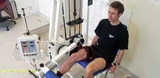
Popularised in the mid-1980s in Japan by Yoshiaki Sato, Blood flow restriction training simply manipulates the normal interaction of motor command, muscle activation, blood supply, fascia movement and nerves fibre communication. The restriction of blood flow is achieved usually in research by specialised pressure cuffs placed at the top of a limb which are inflated to a set pressure throughout exercise. The pressure applied should be high enough to occlude venous return from the muscle but low enough to maintain arterial inflow into the muscle. Venous pooling occurs and local arterial hypoxia stimulates increased blood flow to the area. The literature shows that the end result of such manipulation is an improvement in strength and hypertrophy with the use of significantly lighter loads (≤ 30% 1-RM) than when performing conventional hypertrophic or strength (≥ 75% 1-RM) based resistance training. From a rehabilitative standpoint this is quite an attractive prospect, particularly in the early stages of rehabilitation when limb movement may be compromised.
The study I was involved in a number of years ago examined tissue oxygenation and venous return responses to the varying initial restrictive pressure, target restrictive pressure, and thigh composition (Karabulut et al., 2011). The study highlights that if initial pressures are below 40 mm Hg then deflating cuffs in-between sets or exercises would result is escape of the of accumulated blood and an increased amount of clearing of metabolites produced, thus reducing the augmentation of anabolic stimulating by metabolites. What I mean here by initial pressure is how tightly you wrap the cuff around the limb.
This is an important consideration as to how tight the cuffs are prior to inflation potentially accounts for unequivocal findings in the literature. For example, an 8-week training period performing resistance training with BFR compared to low intensity resistance training without BFR, resulted in no difference in changes in muscular strength (Burgomaster et al., 2003). Importantly, the cuff was deflated during the 5-min recovery interval after the third set and then re-inflated to complete the remaining 3 sets of elbow flexor resistance exercises. Thus the initial pressure may have been below that needed to maintain venous pooling and thus pull the plug of the accumulated pool of blood.
So just make sure the cuffs are tight, right?
Well at initial pressures of 70 mm Hg plus it has been reported that feelings of nausea or dizziness is common. This could be down to a number of factors such as activation of nociceptors relaying the pain experienced due to the tightness. Yet it may be due to the impact of venous blood pooling on stroke volume (Iida et al., 2005). Sharp decreases in stroke volume with increasing levels of the applied-pressure and the reductions in stroke volume were compensated for by an increased heart rate. Changes in femoral blood flow and pooling of blood in the thighs were measured using ultrasonography and the authors indicated that application of 200 mm Hg resulted in decreases in stroke volume of 31.1% due to blood pooling in the vascular and extra-cellular compartment of the legs.
To circumvent the pain and bruising sometimes experienced with relatively high initial pressures (above 70 mm Hg) and the escape of venous pooling from relative low pressures (below 40 mmhg), the suggestion from a recent opinion paper are in line with our suggestions that it would beneficial for future research and for practitioners to set the initial pressure to 50 mm Hg (Loenneke et al., 2013).
Setting the initial pressure is probably only measurable when using the KAATSU system, which is what we used. Using blood pressure cuffs, elastic cuffs or string will definitely occlude. But whether occluding enough, or too much cannot be known. Therefore they can potentially be used but not with the same level of confidence. When a better understanding of the relationship between different initial pressures and subjective responses to such setting the use of anything other than a KAATSU is always going to be second best. More importantly, using methods that cannot account for initial pressure may not only decrease the effectiveness of the intervention, but it may also become a safety concern.
Upon setting an initial pressure of 50 mm Hg, the target pressures often used within the literature to elicit muscular adaptations are anything from 150 to 200 mm Hg. Although going higher than 150 mm Hg no more effective at increasing intramuscular metabolites (Loenneke et al., 2013). Some researchers have based pressure on a percentage of arterial occlusion pressure. Since the goal of BFR is venous pooling without arterial occlusion, the researchers would then take a percentage of this arterial occlusion pressure as the BFR pressure to use for that individual. While this may be an effective means to set a relative pressure with wider cuffs (13.5 cm cuff), the usefulness of this technique with a smaller cuff is questionable as arterial blood flow may not be able to be occluded with a smaller cuff. Thus, basing the pressures on the individuals thigh circumference. This method is likely imperfect but does appear to provide a relative BFR stimulus (Loenneke et al., 2013).
What role does BFR have in rehabilitation and pain management?
On physiological level at the peripheral vasculature, BFR potentiates the expression of a number of important chemical signals that may be really important for improved muscular function. This potentially does not occur during periods of immobilisation or severely reduced activity due to injury.
As I see it there are two main valid reasons as to why BFR would be a useful addition to a Sports Therapist and/or Physiotherapists tool kit, 1) allows for avenue for improvement in strength under compromised movement, 2) may potentially enhance the time taken to return muscle size back to that of pre-injury size.
Evidence suggests the effectiveness of quadriceps strengthening exercises in the treatment of patellofemoral pain, however it is often difficult for patients to perform these exercises at a sufficient load to increase strength (70% of 1 RM) as the high loads may aggravate their patellofemoral symptoms (Chui et al., 2012). Indeed, Hylden and colleagues (2015) have recently shown to improve strength in patients with severe musculoskeletal trauma, persistent chronic quadriceps and hamstring weakness despite traditional therapy, and low improvement during early postoperative strengthening. Patients underwent 2 weeks of BFR training using a pneumatic tourniquet set at 110mmHg while performing low load resistance exercises. Therefore BFR may be used indirectly manage symptoms of pain through improving strength.
To substantiate my second reason low intensity exercise supplemented with BFR has been shown to be an efficient and effective means of maintaining post-surgical muscle size and subjective knee function (Takarada et al., 2000; Ohta et al., 2003; Lejkowski and Pajaczkowski, 2011). Takarada and colleagues (2003) investigated the effect of occlusive stimulus on thigh muscles of patients subjected to the surgical reconstruction of the anterior cruciate ligament (ACL) to see whether it has an effect in diminishing the post-operation muscular atrophy without any exercise stimulus combined. Two sessions of occlusive stimulus, each consisting of five repetitions of vascular occlusion (mean maximal pressure, 238 mm Hg) for 5 min and the release of occlusion for 3 min, were applied daily to the proximal end of the thigh from 3rd to 14th days after the operation. The relative decrease in cross sectional area of knee extensors was significantly larger in the control group (20.7%) than in the experimental group (9.4%). This is good evidence for the use of BFR in a rehabilitative setting.
Conclusion…..
Hopefully I have managed to briefly paint the picture that setting initial pressures are important in being confident of vascular manipulation. In addition, BFR has a potentially important role in rehabilitation from a strength-induced pain relief and hypertrophic perspective. Setting initial pressures of around 50 mm Hg, a target pressure of 150 mm Hg and using loads of 20-30% 1RM would appear to be best practice from the what data is showing us. However there is still so much more to know. I think this area of research and practice would be greatly informed by practitioners partnering up with researchers/scientists to write up the use of BFR in rehabilitative settings as case studies when possible.
Initially, it would probable bear fruit for studies to assess the relationship between objective and subjective measures of pressure due to the practicalities and cost the KAATSU. Though the bottom line is there is not an agreed upon set of guidelines for the use of BFR for rehabilitation purposes thus more work is certainly warranted.
Thanks so much to James for writing this guest blog on a particular area that appears to have some real effectiveness in a rehabilitation setting. There are several clinicians looking into BFR at the present moment and it will be interesting to see what the outcome of their research will be.
Your comments are welcome and as always thanks for reading
TNP
You can find James on twitter: @JPIMcCarron23
Further Reading
Chiu, J.K., Wong, Y.M., Yung, P.S., and Ng, G.Y. (2012). The effects of quadriceps strengthening on pain, function, and patellofemoral joint contact area in persons with patellofemoral pain. American Journal of Physical Medicine & Rehabilitation, 91(2):98-106.
Hylden, C., Burns, T., Stinner, D., and Owens, J. (2015). Blood flow restriction rehabilitation for extremity weakness: a case series. Special Operations Medical Journal, 15(1): 50-6.
Karabulut, M., McCarron, J., Abe, T., Sato, Y., and Bemben, M. (2011). The effects of different initial restrictive pressures used to reduce blood flow and thigh composition on tissue oxygenation of the quadriceps. Journal of Sports Sciences, 29(9):951-8.
Lejkowski, P.M., and Pajaczkowski, J.A. (2011). Utilization of Vascular Restriction Training in post-surgical knee rehabilitation: a case report and introduction to an under-reported training technique. Journal of the Canadian Chiropractic Association, 55(4): 280–287.
Loenneke, J.P., Wilson, J.M., Marin, P.J., Zourdos, M.C., and Bemben, M.G. (2011). Low intensity blood flow restriction training: a meta-analysis. European Journal of Applied Physiology, 112(5):1849-59.
Loenneke, J.P., Fahs, C.A., Rossow, L.M., et al. (2013). Blood flow restriction pressure recommendations: a tale of two cuffs. Frontiers in Physiology, 4: 249.
Takarada, Y., Takazawa, H., and Ishii, N. (2000). Applications of vascular occlusion diminish disuse atrophy of knee extensor muscles. Medicine and Science in Sports and Exercise, 32(12): 2035–2039.

1 Comment