

I’ve got a bit of a habit of doing things backwards. I read magazines from the back to front cause I’m left handed, I tend to act first and think second, I don’t think I’ve ever followed a full physiotherapy assessment by the order of the book, and even my reasoning I’ll experiment with things based upon what the patient tells me at the time. Which, in my opinion is the beauty of adopting an abductive reasoning approach to my practice. For more on that read my previous blog here.
Anyway about doing things backwards. This is just a short reflective blog on a recent experience I had with a patient that has been coming to see me with frozen shoulder (adhesive capsulitis). The patient presented with a typical frozen shoulder pattern – significant painful loss of shoulder range of movement in active and passive ranges. The patient had a normal looking x-ray and recently had a corticosteroid injection which provided no improvement in pain and movement.
I, along with other physio colleagues, am not one for sticking to the rule book or procedure as this usually fails to work. Instead, I like to think outside the box (because it’s interesting and not boring like doing mobilisations, massage and/or stretching which doesn’t really work). I have been attempting eccentric work on frozen shoulders as from my reading of various papers and blogs the way a capsule is formed is not separated from other tissue. It’s kinda all merged into a connective tissue meat market like a Devonshire cattle farm, a mix of eager farmers with west country accents, and a whole bunch of cows all moving in synchrony through the stocks. Anyway, my point is I understand that eccentric training on tendons has physiological effects on the physical make-up and as such increases connective tissue strength and flexibility (O’Sullivan, McAulliffe, & DeBurca, 2014).
So, as my patient and I were attempting to do eccentric training on her shoulder, she noticed something rather odd that she had not recognized before and so did not mention. The patient was lying halfway between her back and right side with the aim of eccentrically lowering her arm into external rotation towards a soft cube, which was used as a marker of progress. She closed her eyes and slowly started to move her arm into external rotation. As she opened them she reported with a surprise “I though my arm was much further away than I thought!” She had managed to get to from internal rotation on her tummy to 0° external rotation when she had perceived her arm to be around 20° external rotation. MISMATCH! I thought immediately. Excitedly, I decided to try a bit of mirror therapy with her to see if we could alleviate (or perhaps elicit) her pain and gain some range of movement. This where things got really interesting.
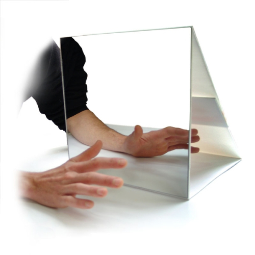
As we were practicing and I was explaining the concept of mirror therapy (watching the reflected image of the asymptomatic arm in the mirror with a view to reduce pain in the symptomatic side), my patient interjected and said “but I already know that my arm is restricted, so how can I be manipulating my brain?” I stood perplexed for a moment and thought actually she has a very good point. The illusion of mirror therapy is to manipulate the brain’s preference of visual feedback over proprioceptive/somatosensory feedback (Moseley, Gallace, & Spence, 2008). Now not being a neuroscientist by any means I felt that the patient had a point that yes if she knows that her arm is restricted then how will a mirror actually convince her otherwise?
So, we decided to flip the mirror round the other way so she would now see the reflected image of her symptomatic arm. We decided to get her to move her asymptomatic side in time with her symptomatic side whilst looking in the mirror. She then would move her asymptomatic side higher (to around 140°) than the reflected image and weirdly she was able to flex her shoulder to 90° with a reduction of pain by about 4 points on a VAS from a 7/10 to a 3/10. It seemed that by flipping the mirror round and watching the reflected image of her symptomatic side whilst moving her healthy shoulder through its full range she recognized that what she saw didn’t match what was normal movement and so implicitly this altered her perception of pain in her symptomatic shoulder. It didn’t improve her movement but it certainly made her more comfortable. All very interesting!
Although a single observational case, it would seem that there was a somatosensory preference over visual feedback when the hidden (asymptomatic) limb was not moved through it’s normal range. It will be interesting to see how her symptoms pan out, the patient has her homework to do so I will feedback towards the end of her pain program.
I have a few questions from this:
- Does the brain always have a preference of visual feedback over proprioceptive/somatosensory feedback?
Perhaps there is equal weighting as we would not expect a non-amputee to present with smudging (or neural migration in the brain) in the same way as an amputee.
- Is this visual feedback preference only apparent in people with phantom limb pain?
This would perhaps make sense as there is no longer proprioceptive/somatosensory information from the amputated limb
- What will the carryover be? How much of the training will have to be explicit in order to maintain change?
- Is this a random case and as such would not be widely reported?
- Can it be used as an analgesic following eccentric training or in combination with eccentric training?
- Could this work for Complex Regional Pain Syndrome?
I’m always keen to get people’s feedback/thoughts.
Thanks for having a read
TNP
References
Moseley, G. L., Gallace, A., & Spence, C. (2008). Is mirror therapy all it is cracked up to be? Current evidence and future directions. Pain. https://doi.org/10.1016/j.pain.2008.06.026
O’Sullivan, K., McAulliffe, S., & DeBurca, N. (2014). the Effects of Eccentric Training on Lower Limb Flexibility: a Systematic Review. British Journal of Sports Medicine, 48(7), 648.2-648. https://doi.org/10.1136/bjsports-2014-093494.234

Leave a Reply